November 17, 2007
Scansion 2
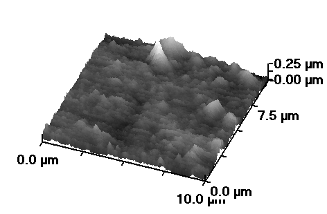
 This image is not, in all honesty, terribly exciting except insofar as it is one of the first scanning ion conductance microscope images I've produced -- indeed one of very few images produced at all on the SICM rig in our lab, which we're just starting to get to grips with.
The scan shows part of the surface of a cultured cell from a line known as AtT20, which is not of any long term interest to my project but is conveniently available for scanning at the moment. Or possibly it shows a grubby patch of dish adjacent to such a cell. Either way, its usefulness is primarily in the acquisition of experience rather than meaningful data.
Still, it's a landmark of sorts.
Posted by matt at November 17, 2007 04:09 PM
Comments
It's a little more abstract than my electron microscope picture of stomatal cells - I just see the peaks of the Himalaya poking above vast cloud banks :)p
Posted by: Alastair at November 18, 2007 09:19 AM
So what's the spiky bit?
Posted by: Anyhoo at November 18, 2007 10:22 PM
No idea. Gunk. I'd point out that the vertical scale is rather different from the horizontal, so it's not nearly as spiky as it looks. Otherwise, that sort of detail will have to wait until I've got other techniques running alongside to help identify what's actually going on down there.
Posted by: matt at November 20, 2007 05:43 PM
Something to say? Click here.


